
HEALTH AND FITNESS
Digital Implant Workflows in 2026: A Practical Guide from Scan to Smile

Key Takeaways
- Digital implant care is strongest when diagnostic records, planning, surgery, and restoration design are connected.
- Accurate scans and complete clinical records help reduce avoidable planning errors.
- Technology supports clinical judgment, but never replaces it.
- Clear communication among the restorative dentist, surgeon, laboratory, and patient is essential.
- Every workflow needs quality checks, trained team members, and a backup plan.
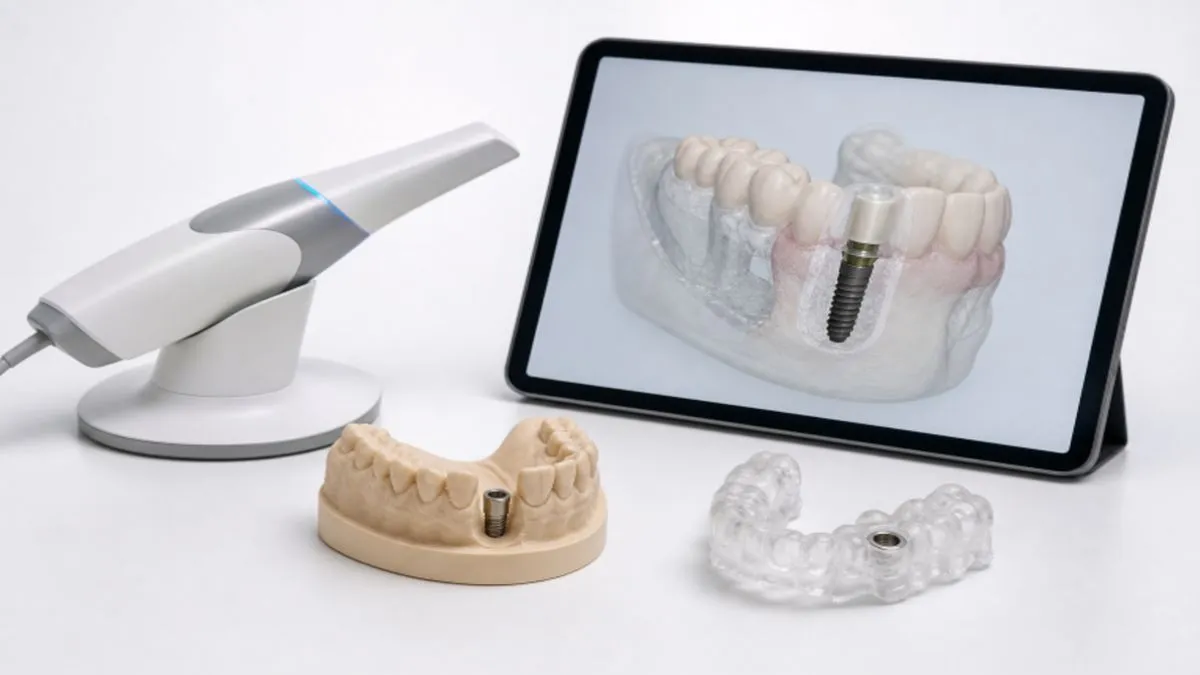
Digital implant dentistry can integrate diagnosis, restorative planning, surgery, laboratory communication, and patient education into a single, more visible workflow. For clinicians building confidence with these systems, Online Dental Implant Courses can provide structured education on the clinical principles behind implant treatment, not just the software steps. The goal is not to make implant care dependent on technology. The goal is to create a reliable process in which every record, decision, and handoff supports the final restoration and the patient’s long-term health.
Table of Contents
Why Digital Workflows Matter in 2026
Modern implant workflows may combine cone beam imaging, intraoral scans, facial records, digital bite information, virtual wax-ups, surgical guides, and CAD/CAM restorations. When these records are reviewed together, the team can visualize the intended tooth position before deciding where to place an implant. This connected approach can make complex cases easier to discuss, document, and transfer between offices or laboratories. It also creates risks when records are incomplete, scans are distorted, files are mismatched, or an operator accepts a digital design without comparing it to the patient’s actual anatomy.
Step One: Start With a Complete Patient Record
Digital planning begins with conventional clinical responsibility. Review medical and dental history, medications, previous treatment, oral hygiene, periodontal condition, occlusion, parafunctional habits, soft tissue, bone condition, and the patient’s priorities. Smoking, uncontrolled diabetes, active periodontal disease, and poor plaque control may affect healing and should be addressed before treatment moves forward. Photographs, diagnostic notes, periodontal findings, and a clear explanation of the restorative goal give the digital records useful context. A detailed scan cannot compensate for an incomplete diagnosis.
Step Two: Combine the Right Digital Records
The best record set depends on the case, but implant teams commonly combine the following information:
- Three-dimensional imaging to assess bone volume and nearby structures, including nerves, sinus spaces, roots, and adjacent implants.
- Intraoral scans to capture teeth, soft tissues, existing restorations, and the planned restorative space.
- Photographs or facial scans to support esthetic planning and communicate visible concerns.
- Digital bite records to evaluate occlusion and available restorative clearance.
- Wax-ups, provisionals, or existing designs when they help define the desired tooth form and position.
Step Three: Plan From the Final Restoration Backward
Restoration-driven planning starts with the proposed crown, not the implant fixture. The team should assess tooth position, emergence profile, esthetics, phonetics, hygiene access, occlusion, material thickness, and the patient’s functional needs. The implant position is then evaluated relative to that intended restoration and the available anatomy. This review should determine whether grafting, soft-tissue management, orthodontic treatment, or another restorative approach may be needed. It should also confirm safe distances from critical anatomy. A technically placeable implant is not automatically a restoratively ideal implant.
Step Four: Build a Team Review Before Surgery
A brief pre-surgical review can prevent expensive misunderstandings. The restorative dentist confirms the tooth design and occlusal plan. The surgeon reviews access, anatomy, bone, guide support, and surgical risks. The laboratory evaluates material choices and manufacturing limits. The coordinator confirms scheduling, consent, and preoperative instructions. Record unanswered questions before approving the final plan. One approved file with a clear version name and date is safer than several similar files circulating via email or messaging systems.
Step Five: Use Surgical Guides with Care
A surgical guide is a transfer device, not a guarantee of success. Before surgery, confirm its fit, seating, stability, support, sleeve design, access, and visibility. The guide must match the patient’s current teeth and anatomy, particularly if teeth have moved, provisional restorations have changed, or records were taken weeks earlier. If a guide does not seat completely, stop and investigate. Do not force it into position or assume the discrepancy is minor. The surgical team should have a documented backup approach for changing anatomy, poor guide fit, limited access, or an unexpected intraoperative finding.
Step Six: Protect Data Quality at Every Stage
A practical quality-control checklist
- Confirm that every image, scan, and design belongs to the correct patient and treatment site.
- Inspect scans for holes, stitching errors, saliva interference, movement, and soft-tissue distortion.
- Compare the digital plan with clinical findings, photographs, and radiographic anatomy.
- Use consistent file names and keep approved files separate from drafts.
- Back up records and limit access to protected patient information.
- Verify laboratory prescriptions, implant components, and restoration details before manufacturing.
What Research Says About Digital Implant Planning
Digital workflows can improve visualization, communication, and procedural consistency, but accuracy still depends on scan quality, planning decisions, guide fit, case selection, and execution. Teams reviewing research on implant placement accuracy should interpret technical deviation data carefully. Placement accuracy is important, but it is only one measure of a successful implant restoration. Recent discussion of digital implant dentistry research also emphasizes that digital tools should be judged by their contribution to patient-centered care, communication, sound decisions, and long-term outcomes, rather than by automation alone.
Improve Patient Communication
Digital images can make implant treatment easier for patients to understand. Show the proposed tooth position, bone limitations, implant location, and expected sequence of care in plain language. Digital previews should be presented as planning aids, not promises that the final result will look exactly like a simulation. Give patients time to ask questions about alternatives, risks, healing, maintenance, cost, and realistic esthetic outcomes. Document informed consent and confirm that the patient understands the limits and benefits of treatment.
Common Problems and Practical Fixes
- Incomplete or distorted scan: Retake the scan and correct the capture process before planning.
- Guide does not seat correctly: Stop, inspect the guide and records, and reassess patient anatomy.
- Plan does not support the final restoration: Return to the restorative design and consult the laboratory.
- Different team members use different files: Establish one approved case file and version-control process.
- Patient expectations are unrealistic: Explain what guided surgery and digital previews can, and cannot, predict.
Where Artificial Intelligence May Fit
AI may assist with image review, record organization, documentation, and patient communication. However, automated suggestions must be checked against original images, clinical findings, and the patient’s specific needs. Teams should also consider consent, data security, privacy, transparency, and who remains accountable for the final clinical decision.
A Simple Implementation Plan for Dental Teams
- Begin with one predictable case type rather than every implant indication.
- Map each step from consultation through final restoration.
- Assign responsibility for scanning, planning, approvals, and file transfer.
- Create a short quality-control checklist and a guide-failure backup plan.
- Review completed cases as a team and document what should change.
- Add complexity only after the workflow is consistent and the team is trained.
Conclusion
A robust digital implant workflow is defined by careful documentation, restoration-driven planning, reliable communication, and clinical judgment. The newest scanner or software platform cannot replace those fundamentals. When technology is used as part of a disciplined care system, it can help dental teams move from scan to smile with greater clarity, consistency, and focus on the patient. A successful approach combines accurate diagnostics, efficient collaboration between specialists and laboratories, and thoughtful treatment planning at every stage. By balancing innovation with proven clinical practices, dental professionals can improve predictability, reduce unnecessary complications, and create a smoother experience for both providers and patients. The goal is not simply adopting new tools, but using them effectively to support better outcomes.


Insetprag: A New Paradigm in Context-Aware System Design

Explore Coomersu: How Community and Commerce Create a Unique Shopping Experience

OnionPlay: The Shocking Truth About Free Streaming

Kaliscan: Read Free Manhua & Manhwa Comics Online

Digital Implant Workflows in 2026: A Practical Guide from Scan to Smile

CHAS6D Explained: Cybernetic Systems in Six Dimensions

Assimilasjon: A Complete Guide to Its Dimensions

Discretion by Design: How Private Households Vet Everyone They Hire

Insanony: The Ultimate Tool for Anonymous Instagram Story Viewing

The Jacksonville Computer Network Issue: A Comprehensive Analysis

How Dermatologists Treat Chronic Skin Conditions

Çrbiti: Symbol of Fluidity, Transformation, and Unpredictability

MonkeyGG2: Explore a World of Games and Socialize with Players

Malaysia Law Firms and Letters of Demand: What You Need to Know

Rowdy Oxford Integris: A Modern Archetype of Edge & Ethics
-
 GENERAL1 year ago
Christofle – For Those Who Dream of Family Heirloom Silver
-
SPORTS3 weeks ago
Discover the World of Football with Streameast: Watch Your Favorite Leagues and Tournaments
-
HOME IMPROVEMENT1 year ago
Spacious and Stylish 300cm Round Rugs: Elegant and Modern Designs for a Chic Home Decor
-
GENERAL8 months ago
Uncovering the World of кинокрадко: The Dark Side of Film Piracy